優秀論文
張明華1,朱曼1,陳超1,裴斐1,石莉1,孫艷2,*,郭代紅2,*
1中國人民解放軍總醫院,藥品保障中心,臨床藥學室,100853
2中國人民解放軍總醫院,藥品保障中心,100853
*通訊作者: guodh301@163.com 或者 301sunyan@sina.cn
摘要
MLN4924是一種選擇性的NAE抑制劑,希望成為腫瘤治療藥物。MLN4924能夠抑制Cullin-RING泛素連接酶活性,上調其底物,干擾腫瘤細胞的S期DNA合成,最終導致腫瘤細胞的凋亡。Cullin-RING的泛素連接酶活性需要Neddylation體系的修飾。HECT類泛素連接酶ITCH在腫瘤的發展過程中具有重要的作用,可能與Neddylation也有密切的關系。探索ITCH的泛素連接酶作用機制就可能有助于更全面地理解MLN4924的治療機制。我們發現,Nedd8的修飾可激活ITCH的泛素連接酶活性,促進對p73等底物的降解,其活性可被MLN4924抑制。ITCH在結直腸腫瘤中顯著高表達。敲低ITCH后結直腸癌細胞系的增殖和遷移能力受到顯著的抑制,且與MLN4924存在協同作用。ITCH的激活機制及其在腫瘤中的調控作用使其有可能成為新的腫瘤藥物作用靶點。
關鍵詞:蛋白質降解,泛素連接酶, Neddylation,ITCH,結直腸癌
Targeting HECT-type E3 ligase ITCH for Inactivation by MLN4924 in Colorectal Cancer Cell Line
Minghua Zhang1, Man Zhu1, Chao Chen1, Fei Pei1, Li Shi1, Yan Sun2,*, Daihong Guo2,*
1Department of Clinical Pharmacy, Center of Pharmacy, Chinese PLA General Hospital, Beijing 100853, China
2 Center of Pharmacy, Chinese PLA General Hospital, Beijing 100853, China
*Correspondence: guodh301@163.com or 301sunyan@sina.cn
Abstract
MLN4924 is a potent and selective inhibitor of NAE, holding promise for the treatment of cancer. The mechanism of MLN4924 was thought to be disrupting cullin-RING ligase-mediated protein turnover leading to the deregulation of S-phase DNA synthesis and apoptotic death in human tumour. Cullin-RING E3s are activated via modification by the ubiquitin-like protein Nedd8 in a process called Neddylation. Neddylation maybe also involved in the activation of HECT-type E3 ligase ITCH, wich plays an important role in tumorigenicity. So, deciphering the activation mechanisms of ITCH is critical for understanding the mechanism of MLN4924 in cancer treatment. Here, we show that ITCH is activated by Neddylation, promoting its ubiquitination ligase activity and the degredation of p73, wich can be inhibited byMLN4924. ITCH expression is elevated in colorectal cancer tissues compared with adjacent tissues. Further experiments indicated that knockdown of ITCH in HCT116 cells resulted in impaired cell growth and migration. And there is a synergy between shRNA-ITCH and MLN4924. These findings shed new light on the activation of ITCH and its potential role to be an anti-tumour drug target.
Keywords: protein degradation; ubiquitin ligase; Neddylation; ITCH; colorectal cancer
以色列人阿龍·切哈諾沃(AaronCiechanover)發現了泛素蛋白酶體系統,并因此在2004年與阿夫拉姆·赫什科(Avram Hershko)、歐文·羅斯(Irwin Rose)一道獲得諾貝爾化學獎[1]。泛素蛋白酶體系統的降解過程需要三種酶參與。首先,泛素活化酶(E1)消耗ATP,并腺苷酸化一個泛素分子。接著,泛素轉運到E1的活性中心,與此同時,E1腺苷酸化第二個泛素[2]。隨后泛素分子被轉移到泛素結合酶(E2),與E2的半胱氨酸殘基結合。最后,泛素連接酶(E3)將泛素分子從E2轉移到高特異性的蛋白底物上,只有四個泛素分子組成的泛素連結合到底物分子上后,這個底物蛋白才會被蛋白酶體識別和降解[3]。所以,底物的特異性由泛素連接酶決定[4]。
泛素蛋白酶體系統負責調控多種信號通路中關鍵蛋白因子的降解[5]。而且也有越來越多的證據表明泛素蛋白酶體系統參與了腫瘤的發展。腫瘤中泛素連接酶的表達擴增或缺失已經多有報道[6, 7]。很多泛素連接酶也已經被鑒定為癌基因(比如BRCA1和 Fbw7)或抑癌基因(比如 Mdm2 和 Skp2)。在多數結直腸癌中,由于APC失活,導致泛素蛋白酶體降解β-連環蛋白能力下降,從而導致腫瘤細胞增殖失控。Nedd4家族泛素連接酶ITCH與腫瘤的關系也非常密切[8-11]。一些調控腫瘤的重要分子,比如p53,Smad4和k-ras家族的某些成員等,也受到了泛素連接酶的調控。在泛素化反應的體系中,泛素連接酶具有底物識別的特異性,從而有可能成為腫瘤靶向性治療的更好的靶標。
有些分子在結構上和泛素分子比較相似,也能夠象泛素一樣被修飾到底物上,這些分子統稱為類泛素分子。目前已經發現的類泛素分子至少有17種[12]。其中Nedd8 (neural-precursor-cell-expressed developmentally down-regulated 8)是與泛素關系最為密切的一個。它與泛素在一級序列上有80%的相似度,在高級結構上也非常相似[13]。催化Nedd8結合到底物上的一系列酶也和泛素化系統非常類似[14]。能夠被Nedd8修飾的底物分子比泛素要少,研究較多的底物是Cullin類分子。Cullin類分子是CRLs (cullin-RING ubiquitin ligases)類泛素連接酶的支架分子。Nedd8修飾到Cullin類分子上,使其空間構象發生改變,同時阻止Cand1對Cullin的抑制作用[15, 16]。
由千禧年藥業有限公司(Millennium Pharmaceuticals, Inc.)和強生公司聯合開發的多發性骨髓瘤治療藥物Velcade,就是一種蛋白酶體抑制劑,該藥物已經成功用于臨床治療惡性骨髓瘤,而且對慢性淋巴細胞白血病的治療也已經處于臨床實驗階段[17, 18]。Velcade的成功應用提示泛素蛋白酶體系統中的關鍵酶可以作為腫瘤等疾病的靶點,用于開發新的腫瘤靶向性藥物[19, 20]。但是Velcade對泛素蛋白酶體系統的抑制是廣譜的,而不是特異的靶向腫瘤細胞,具有相對比較廣泛的不良反應。千禧年藥業新一代泛素化抑制劑MLN4924進一步明確了作用靶點,縮小了抑制范圍,對結直腸癌獲得了顯著的療效,與Velcade相比,明顯地減少了不良反應[21]。目前認為,MLN4924可能就是通過高效地具有選擇性地抑制Nedd8活化酶(NAE),干擾cullin-RING泛素連接酶介導的蛋白質的轉歸(protein turnover),從而導致腫瘤細胞周期S期DNA合成失調,最終導致腫瘤細胞凋亡。
然而,最近幾年發現,Neddylation和泛素化兩條通路存在更多的交叉[22-24]。并且這種交叉在細胞內比較普遍,比如在各種應激、自由的泛素分子減少時、Nedd8分子增加時(比如Nedd8過表達時)、蛋白酶體受到抑制等條件下,都會發生。在細胞內過表達Nedd8時,甚至會發現,本應完全由泛素組成的泛素鏈,中間的某些泛素分子可能會被Nedd8所取代,成為由泛素和Nedd8共同組成的一條“類泛素蛋白鏈”[22]。還有一些連接酶既是Neddylation系統的E3,又是泛素化系統的E3。所以,Nedd8修飾某些底物之后,作用就很難一概而論,需要在具體通路、具體分子和具體條件下進行分析;MLN4924對腫瘤的作用機制,也就增加了很多可能性。在前期的研究中我們發現,HECT類泛素連接酶ITCH可能就處于兩個通路的交叉點上,其活性也受到MLN4924的調控。
1. 材料與方法
1.1 材料與試劑
Nedd8、ITCH的表達序列通過PCR構建到相應表達載體上。pGEX-4T-2載體購自Amersham公司。pCMV-Myc真核表達載體購自Clontech公司。MLN4924購自Active BioChem公司。Pyrobest DNA Polymerase、DL-2000 DNA marker、dNTP、DL-5000 DNA marker、T4 DNA連接酶和限制性內切酶均購買于TaKaRa公司;逆轉錄試劑盒購買于Toyobo公司;質粒提取試劑盒、凝膠回收試劑盒均購買于Promega公司;RPMI 1640、DMEM、DMEM/F12、胎牛血清及雙抗及胰酶購買于Hyclone公司;感受態細胞BL21、DH5α、GSTK抗體購自TIANGEN;TRIZOL、Lipofectamine2000、FITC標記的羊抗鼠抗體購買于Invitrogen公司;NC膜、尼龍膜購買于Amersham公司;Western blot實驗顯色試劑盒購買于Pierce公司。
1.2 免疫印跡
除去培養液,1ml的PBS洗滌2次。滴入裂解液以及等量的2×loading并混勻。.沸水煮0-15分鐘并1000g離心4分鐘。進行SDS-PAGE,并轉膜。含有5%脫脂牛奶的TBST慢搖1h。封口膜包裹,并于4℃過夜。使用TBST洗三次,每次10min左右。在室溫中孵育二抗45分鐘,并TBST洗滌三次,每次10分鐘左右,加發光底物并進行曝光顯影。
1.3 免疫組化
將白片進行脫蠟:2×濃度的二甲苯15min。 水化的順序如下:100%乙醇 3min,95%乙醇 3min,90%乙醇 3min,80%乙醇 3min;重復步驟b,用蒸餾水沖干凈切片。除去內源過氧化物酶:使用3% H2O2 室溫孵育10min(PBS配置,注意現用現配,隔絕氧),PBS洗2min X 3。 抗原修復:向抗原修復盒中灌滿PBS溶液,在微波爐中中火加熱5min,在PBS將沸未沸時(95度),置入切片,換成小火5min加熱,后取出修復盒,冷卻至室溫后,約需2小時,取出切片。Trition-100破膜:將切片平鋪于冰上,滴上PBS配置的Triton X-100 (0.5%-1%),20min。完畢后PBS清洗洗3遍,血清封閉37℃1小時。一抗孵育:一抗用3% bsa-pbs配置,濕盒中37℃孵育2小時或4度過夜。完畢后PBS洗3遍。使用中山金橋二步法試劑盒。Dab顯色。
1.4 細胞生存和遷移實驗
利用假病毒顆粒感染細胞,獲得穩定株,傳代鋪入24孔板。每隔24h,胰酶消化吹散細胞,取10ul混懸液加入一次性計數板,電子計數。
于Transwell孔中加入無血清培養基,下層培養皿中加入含5%血清的培養基,將穩定株細胞鋪入transwell孔中培養,24小時后取出transwell孔,清洗干凈后甲醇固定30分鐘,結晶紫染色20分鐘,清洗晾干后掃描。
2. 結果
2.1 Nedd8與ITCH存在相互作用,且Nedd8能夠共價修飾ITCH,這種修飾依賴于Neddylation體系。
在結腸癌細胞系HCT116中,我們利用內源的ITCH抗體檢測Nedd8抗體的免疫沉淀樣品,發現Nedd8與ITCH存在相互作用(Fig. 1A)。純外源pull down實驗結果顯示ITCH和Nedd8存在直接的相互作用(Fig. 1B)。在HCT116細胞系中免疫共沉淀后,利用Nedd8抗體檢測共純化的ITCH蛋白,我們發現Nedd8對ITCH的修飾結果為連續的條帶狀,且這種修飾受到MLN4924的抑制(Fig. 1C)。此外,利用siRNA干擾Neddylation E1的亞基Uba3后,Nedd8對ITCH的修飾同樣顯著降低(Fig. 1D)。
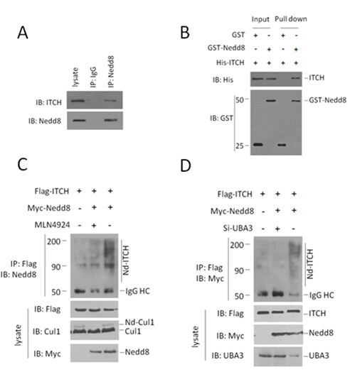
Fig.1 (A) Endogenous interaction of ITCH and Nedd8 in HCT116 cells. Both the cell lysates and immunoprecipitates were analysed by western blotting with the indicated antibodies. (B) GST pull-down assay was performed to detect the direct interaction between ITCH and Nedd8. Both input and pull-down samples were subjected to immunoblotting (IB) with anti-His and anti-GST antibodies. Input represents 10% of that used for pull-down. (C,D) ITCH was neddylated in HCT116 cells. This neddylation was significantly attenuated by NAE inhibitor MLN4924, or depletion of Uba3 (one subunit of NAE). The indicated plasmids or siRNAs were transfected into HCT116 cells. Flag antibody-immunoprecipitated ITCH proteins were analyzed by immunoblotting with anti-Myc antibody to detect conjugated Nedd8.
2.2 Nedd8的修飾促進ITCH對底物的泛素化活性。
ITCH的泛素化底物包含P73、P63、LAPTM5和RASSF5/NORE1等重要的分子,并可能通過這些底物參與腫瘤發展的調控[10, 11, 25]。在HCT116細胞系中過表達外源Nedd8、ITCH、P73和泛素,利用外源P73抗體免疫沉淀后,檢測外源Ub,我們發現,Nedd8可促進ITCH對P73的泛素化水平(Fig . 2A)。同時,Nedd8能夠促進ITCH對P73的降解,而且我們發現與其他HECT類泛素化連接酶類似,ITCH在降解底物時自身降解也在加速(Fig . 2B)。
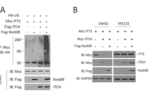
Fig.2 (A) Nedd8 promotes the ubiquitination activity of ITCH in HCT116 cells. The indicated plasmids were transfected into HCT116 cells. Myc antibody immunoprecipitated Ub proteins were analyzed by immunoblotting with anti-HA antibody to detect conjugated Ub. (B) P73, ITCH and Nedd8 were co-transfected into HCT116 cells. Cell lysates were immunoblotted to detect p73 levels.
2.3 ITCH在結直腸腫瘤中顯著高表達,在腫瘤細胞分裂期與中心體和微管存在共定位。
為探索ITCH在結直腸腫瘤中的表達水平,利用免疫組化技術在80例結直腸腫瘤樣本的蠟塊切片中檢測ITCH的表達水平。我們發現,腫瘤組織中ITCH的蛋白水平顯著高于同一患者的癌旁組織(Fig. 3A)。ITCH主要定位于細胞質,且分布不均勻,分裂間期細胞中,ITCH的染色往往聚集成團塊狀,靠近細胞核(Fig. 3B)。為了進一步檢測ITCH的亞細胞定位,利用內源ITCH抗體、α-tublin抗體和DAPI,在Hela細胞中進行激光共聚焦,我們發現,在細胞的分裂期,ITCH主要定位于中心體(Fig. 3C)。
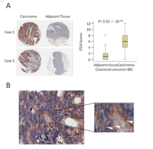
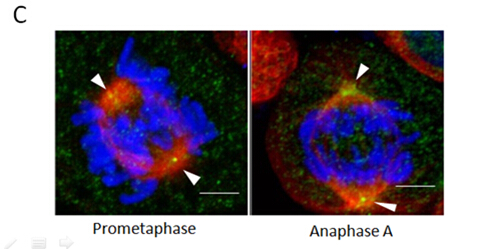
Fig.3 ITCH overexpressed in colorectal cancer and locallized in centrosomes in mitosis. (A) ITCH expression scores are shown as box plots, with the horizontal lines representing the median; the bottom and top of the boxes representing the 25th and 75th percentiles, respectively; and the vertical bars representing the range of data. Colorectal cancer tissues were compared with matched adjacent normal tissues using the Wilcoxon test. n = 80. Representative images from immunohistochemical staining of ITCH expression of the same tumor from three cases. Scale bars, 100um.(B). Colorectal cancer tissue stained with antibodies to the ITCH protein. Bars, (left) 40 um, (right) 20um.(C) Representative images of ITCH protein localization in metaphase. The cells were stained with 4,6-diamidino-2-phenylindole(DAPI; for DNA; blue), anti-tubulin (microtubules; red) and ITCH antibody (kinetochores; green). Scale bar, 10 um.
2.4 ITCH在結直腸腫瘤中顯著高表達,在腫瘤細胞分裂期與中心體和微管存在共定位。
我們發現ITCH在結直腸腫瘤中顯著的高表達,其泛素化活性受到MLN4924的抑制。為了探索靶向ITCH 的shRNA和 MLN4924對腫瘤細胞的發展有何作用,我們利用攜帶ITCH-sh-RNA的慢病毒體顆粒感染HCT116細胞后檢測其增殖和遷移的能力,并檢測在這些細胞系中MLN4924的作用。我們發現,靶向ITCH 的shRNA可以顯著抑制結直腸細胞系的增殖和運動能力,且與MLN4924存在協同作用(Fig. AB)。
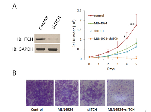
Fig.7 ShRNA targeting ITCH depressed the growth and metastasis of colorectal cancer cell line in vitro. (A)Lentivirus-coupled shRNA ITCH and control shRNA vectors were each stably transfected into HCT116 cells. The expression of ITCH was detected by western blot to show the depletion efficiency (lane 2). *p < 0.01, **p < 0.001 (unpaired two-tail t test). (B) The in vitro cell migration assay was performed in transwell plate. Representative results from three independent experiments are shown, bars, 100um.
3討論
靶向泛素-蛋白酶體系統的抑制劑Velcade,目前已經用于治療多發性骨髓瘤和淋巴瘤,效果良好。同一個公司開發的通過靶向Neddylation過程治療結直腸癌的新藥MLN4924,療效更為顯著,不良反應更小,已經進入II期臨床實驗,很可能成為新一代的重磅炸彈新藥。前期的研究發現MLN4924能夠抑制Culling類的泛素連接酶活性,上調周期調控因子,使腫瘤細胞周期阻滯,促進凋亡,從而達到治療目的[12, 17, 21]。近期有報道Neddylation與Ubiquitination的交叉可能遠比已知的要多,那么MLN4924在抑制Neddylation過程時,就會影響更廣泛的泛素化過程。
我們發現ITCH具有Neddylation連接酶活性,并揭示了Nedd8對ITCH泛素化連接酶活性的調控作用。在結直腸腫瘤中ITCH高水平表達,在分裂期定位于中心體,提示ITCH很可能參與了腫瘤細胞的周期發展和調控。在結直腸腫瘤細胞系中敲低ITCH與MLN4924的作用一致,都能抑制腫瘤細胞的增殖和遷移,且ITCH的干擾RNA可起到MLN4924的協同作用,同時使用,大大增強了對腫瘤細胞的抑制作用。在泛素化反應的體系中,泛素連接酶具有底物識別的特異性,從而有可能成為腫瘤靶向性治療的更好的靶標。因此,我們的研究不僅發現了MLN4924抑制腫瘤發展的一種新機制,而且為設計抗結直腸腫瘤藥物提供了新的潛在藥物靶標。
參考文獻
1. Neefjes J, Groothuis TA, Dantuma NP: [The 2004 Nobel Prize in Chemistry for the discovery of ubiquitin-mediated protein degradation]. Ned Tijdschr Geneeskd 2004, 148(52):2579-2582.
2. Haas AL, Warms JV, Hershko A, Rose IA: Ubiquitin-activating enzyme. Mechanism and role in protein-ubiquitin conjugation. J Biol Chem 1982, 257(5):2543-2548.
3. Thrower JS, Hoffman L, Rechsteiner M, Pickart CM: Recognition of the polyubiquitin proteolytic signal. EMBO J 2000, 19(1):94-102.
4. Risseeuw EP, Daskalchuk TE, Banks TW, Liu E, Cotelesage J, Hellmann H, Estelle M, Somers DE, Crosby WL: Protein interaction analysis of SCF ubiquitin E3 ligase subunits from Arabidopsis. Plant J 2003, 34(6):753-767.
5. Hershko A: The ubiquitin system for protein degradation and some of its roles in the control of the cell division cycle. Cell Death Differ 2005, 12(9):1191-1197.
6. Chen C, Seth AK, Aplin AE: Genetic and expression aberrations of E3 ubiquitin ligases in human breast cancer. Mol Cancer Res 2006, 4(10):695-707.
7. Comprehensive molecular characterization of human colon and rectal cancer. Nature 2012, 487(7407):330-337.
8. Oberst A, Rossi M, Salomoni P, Pandolfi PP, Oren M, Melino G, Bernassola F: Regulation of the p73 protein stability and degradation. Biochem Biophys Res Commun 2005, 331(3):707-712.
9. Salah Z, Melino G, Aqeilan RI: Negative regulation of the Hippo pathway by E3 ubiquitin ligase ITCH is sufficient to promote tumorigenicity. Cancer Res 2011, 71(5):2010-2020.
10. Rossi M, De Simone M, Pollice A, Santoro R, La Mantia G, Guerrini L, Calabro V: Itch/AIP4 associates with and promotes p63 protein degradation. Cell Cycle 2006, 5(16):1816-1822.
11. Suryaraja R, Anitha M, Anbarasu K, Kumari G, Mahalingam S: The E3 ubiquitin ligase Itch regulates tumor suppressor protein RASSF5/NORE1 stability in an acetylation-dependent manner. Cell Death Dis 2013, 4:e565.
12. Schulman BA, Harper JW: Ubiquitin-like protein activation by E1 enzymes: the apex for downstream signalling pathways. Nat Rev Mol Cell Biol 2009, 10(5):319-331.
13. Whitby FG, Xia G, Pickart CM, Hill CP: Crystal structure of the human ubiquitin-like protein NEDD8 and interactions with ubiquitin pathway enzymes. J Biol Chem 1998, 273(52):34983-34991.
14. Gong L, Yeh ET: Identification of the activating and conjugating enzymes of the NEDD8 conjugation pathway. J Biol Chem 1999, 274(17):12036-12042.
15. Liu J, Furukawa M, Matsumoto T, Xiong Y: NEDD8 modification of CUL1 dissociates p120(CAND1), an inhibitor of CUL1-SKP1 binding and SCF ligases. Mol Cell 2002, 10(6):1511-1518.
16. Zheng J, Yang X, Harrell JM, Ryzhikov S, Shim EH, Lykke-Andersen K, Wei N, Sun H, Kobayashi R, Zhang H: CAND1 binds to unneddylated CUL1 and regulates the formation of SCF ubiquitin E3 ligase complex. Mol Cell 2002, 10(6):1519-1526.
17. Yang Y, Kitagaki J, Wang H, Hou DX, Perantoni AO: Targeting the ubiquitin-proteasome system for cancer therapy. Cancer Sci 2009, 100(1):24-28.
18. Eldridge AG, O'Brien T: Therapeutic strategies within the ubiquitin proteasome system. Cell Death Differ 2010, 17(1):4-13.
19. Berkers CR, Ovaa H: Drug discovery and assay development in the ubiquitin-proteasome system. Biochem Soc Trans 2010, 38(Pt 1):14-20.
20. Nalepa G, Rolfe M, Harper JW: Drug discovery in the ubiquitin-proteasome system. Nat Rev Drug Discov 2006, 5(7):596-613.
21. Soucy TA, Smith PG, Milhollen MA, Berger AJ, Gavin JM, Adhikari S, Brownell JE, Burke KE, Cardin DP, Critchley S et al: An inhibitor of NEDD8-activating enzyme as a new approach to treat cancer. Nature 2009, 458(7239):732-736.
22. Leidecker O, Matic I, Mahata B, Pion E, Xirodimas DP: The ubiquitin E1 enzyme Ube1 mediates NEDD8 activation under diverse stress conditions. Cell Cycle 2012, 11(6):1142-1150.
23. Hjerpe R, Thomas Y, Kurz T: NEDD8 overexpression results in neddylation of ubiquitin substrates by the ubiquitin pathway. J Mol Biol 2012, 421(1):27-29.
24. Hjerpe R, Thomas Y, Chen J, Zemla A, Curran S, Shpiro N, Dick LR, Kurz T: Changes in the ratio of free NEDD8 to ubiquitin triggers NEDDylation by ubiquitin enzymes. Biochem J 2012, 441(3):927-936.
25. Ishihara T, Inoue J, Kozaki K, Imoto I, Inazawa J: HECT-type ubiquitin ligase ITCH targets lysosomal-associated protein multispanning transmembrane 5 (LAPTM5) and prevents LAPTM5-mediated cell death. J Biol Chem 2011, 286(51):44086-44094.
北京藥學會 地址:北京市朝陽區北三環中路2號小二樓2層
本網站瀏覽46593654次
Copyright 2012 北京藥學會( 本網站所有內容未經許可,不得以任何形式進行轉載 ) All Rights Reservered
